

Telling the difference between Mycoplasma, Quantum Dots and Chylomicrons
Telling the difference between Mycoplasma, Quantum Dots and Chylomicrons

There was a question about how to tell the difference of these in Live Blood Analysis. This is how I was trained for mycoplasma and chylomicrons, we were NOT trained in quantum dots in the blood, obviously, since no one knew of them until now. I am sure there may be different opinions.
The first clue is what are you looking at, bright field or dark field. I also feel that you have to look at all three methods, bright field, dark field and dried blood to really know and understand what is happening to someone’s blood.
Chylomicrons are anomalies of platelets and plasma. They are the largest and least dense of all lipoproteins and consist of 85% triglycerides, 7% phopholipids, 6-7% cholesterol and 1-2% protein. Chylomicrons are the only lipoproteins that are a reflection of diet. So inquiring what the person eats and how long it’s been since they have eaten is really important.
I rarely see chylomicrons as I always ask the patient to be fasting for 4 hours prior to drawing their blood. Chylomicrons are viewed in 2 phases, either delayed appearance or delayed clearance. Chylomicrons are viewed in Dark Field and are not easily seen in bright field. The images of mycoplasma that I showed in the last post were of Bright field. I have rarely seen chylomicrons in bright field.

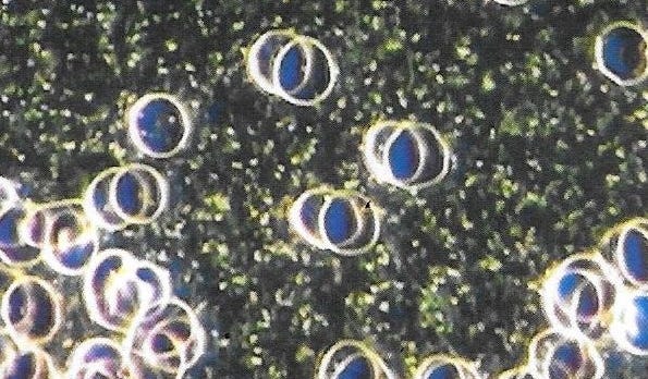
In delayed appearance of chylomicrons very few will be observed in someone who consumed a meal containing fat within 2-3 hours of being tested. Here is a dark field view of chylomicrons delayed appearance;
In chylomicrons delayed clearance are small white dots, moving around actively and again not easily seen in bright field. Again knowing whether they are in a fasting state or 4 hours after a meal is important to determine if it’s delayed clearance.
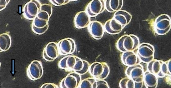
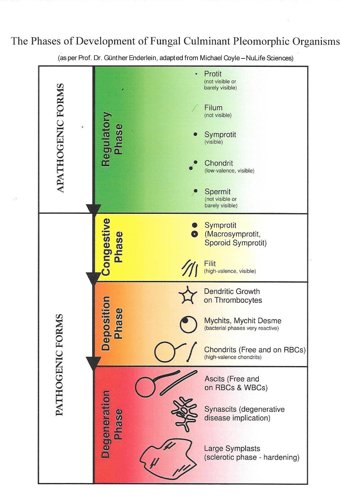
Mycoplasma are small, spheres or donut shaped forms that can be seen moving around actively in the plasma or attached to RBC’s. They can only be easily viewed in bright field and confusing somewhat in dark field because they are synonymous with sporoid symprotits and macrosymprotits (see chart below this next image) in pleomorphism which is the philosophy that live blood analysis follows. Here is a comparison of bright field and dark field of mycoplasma;
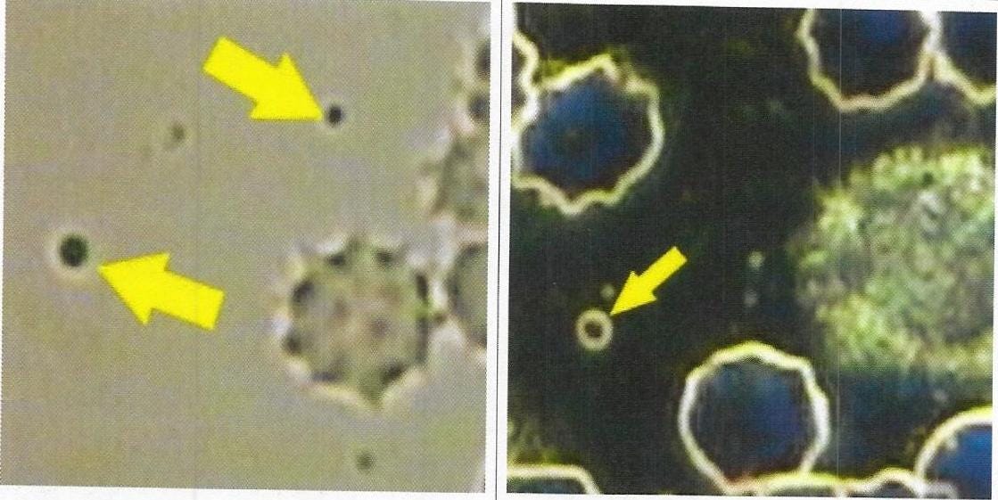
Chart of pleomorphism:
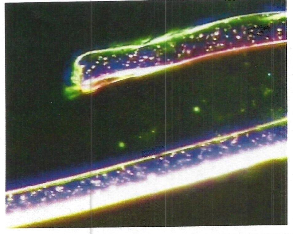
In my opinion the difference between mycoplasma and chylomicrons above and quantum dots in the blood is the color of them. Red or pinkish, green or blue. Here are some images that show these colorful little dots;


We have to be careful not to confuse what we are seeing with artifacts like foreign objects from a microfiber lens cloths that might be used to wipe off the slides like this;
Obviously none of the images that I have posted here were left overnight like many are doing to see what develops, I am only trying to show what I believe the differences are of mycoplasma, chylomicrons and quantum dots. Take care.
Hi Paulette.
Hi Paulette.
Could I send you some enlarged images by email and could you clarify if what is observed is mycoplasma?