
Validity of Darkfield Microscopy

There are many negative comments out there in Internet Land about Live Blood Analysis. Most of them say that we, Certified Live Blood Analysts are diagnosing disease with Live Blood viewed with a Darkfield Microscope. We are not diagnosing disease, Certified Practitioners know that diagnosing disease is only for MD’s. However, as a Certified Live Blood Analysts, I can understand why there is so much confusion about Darkfield Microscopy.
I have been practicing Live Blood since 2018. I have taken the certification course and an advanced course which was completed in 2021. In our training we learn about artifacts which are caused from incorrect sampling techniques, cheap slides or cover slips, squeezing the finger, alcohol not wiped off before sampling, foreign materials like fibers and air bubbles, which are hard to avoid.
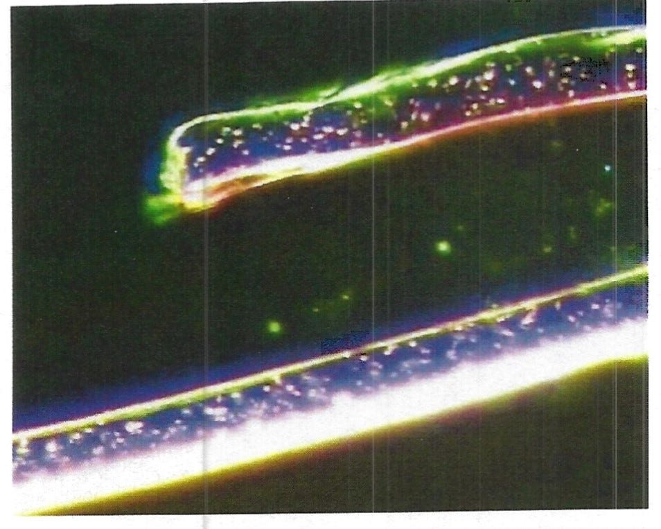
This might look familiar, like the hydrogel filaments we have been seeing in blood. I assure you the hydrogel filaments are real, but this is a picture of a fiber from a lens cloth. This is why I stopped using them. (If you can’t see the picture, click on the title of this article)
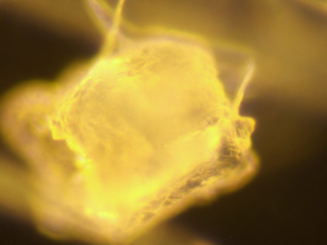
How about this? Sort of looks like the chips we have been seeing being formed in the blood, right? No, this is a fragment from a lens cloth.
I assure you I am not saying that the hydrogel filaments we have been seeing or microchips being formed in our blood are not real, they are real. I am trying to show why there is so much controversy about Live Blood Analysis.
Another area that is a problem is Rouleau. The blood cells that looked like a stack of coins.
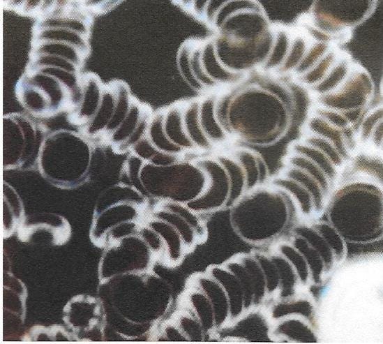
This can be created by squeezing the finger when trying to get the sample. When I get this finding with a patient, I immediately ask to take another sample. I just have to be sure that I didn’t make the mistake, if after careful sampling again, the Rouleau is there, then it is an accurate finding of sticky blood.
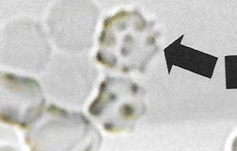
Another finding from squeezing the finger during sampling looks like Echinocytes or Poikilocytes. Echinocytes are very spiky, like this:
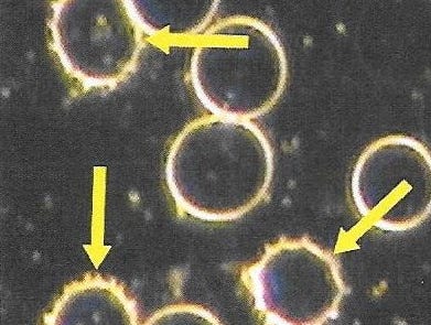
Poikilocytes look like these cells with dots all over them:
Both of these anomalies can be in every sample especially at the edges of the coverslip. You see there is a working area of a sample, we avoid the zone of aggregation, again the edges of the coverslip where the blood is thick, you will see clumping of the cells and we avoid the area called zone of Lysis, where there is almost no blood cells. But if the anomaly does show in the working area, then we make recommendations of detoxification.
This doesn’t mean we don’t look at the whole sample, we do, we just don’t make recommendations based on those areas, only the working area. With all the hydrogel stuff we are seeing, we look at the whole sample.
Some brilliant darkfield practitioners like
, have been showing interesting items showing up in the air bubbles. We darkfield practitioners don’t make recommendations based on the air bubbles but Matt and Karl are showing the hydrogel filaments looking like the air bubble’s are creating them. This is all so unknown, we are all learning here.So I say to the naysayers out there who say that darkfield microscopy is not valid, how come they use it in pathology? In pathology they use it to see spirochetes for disease, how come that is valid?
Is dark-field microscopy still useful for the primary syphilis diagnosis in the 21ST century? https://doi.org/10.1016/j.eimce.2021.10.001
Conclusions
DFM allows primary syphilis early diagnosis, even without serological test.
They see the squiggly little spirochetes moving around,

why is it that when we practitioners see bacteria, or fermentation, thrombosis, fibrin, that it is not valid? For some reason they feel it’s only valid with tissue not blood. There has to be some common sense here, really!
Why do they teach it in Universities, if it’s not valid.
Essentials of Medical Microbiology First Edition: 2016 ISBN: 978·93·5 152s987·3
They clearly say in this text book, “Applications; Dark field microscope is used to identify the living. unstained cells and thin bacteria like spirochetes which cannot be visualized by light microscopy.”
So we, Darkfield Microscopy practitioners, know better! It is a valid way of looking at live blood but the practitioner must be careful in taking the sample.
Thank you for this! I just got a microscope and I’m trying to figure out what I’m looking at. I had a ton of spiky RBC’s. I did squeeze my finger so I assume that’s why I had a portion of the sample very spiky.
What school certified you in DFM? Thank you for this. I've been interpreting my blood incorrectly.